|
GALLERY OF IN VIVO EMBRYO DEVELOPMENT (ALL MOVIES, click on the links to get the time-lapse animations)
(NB : the french page is updated more frequently)
Last update Sanuary 2015.
Last update September 2012.
The movies span all stages between a formless mass of cells, and a recognizable vertebrate, having a head, vertebrae precursors etc.
All video movies are available in 768x512 format. In some cases HD movies are also available in 1200x746. Several movies are dual video films taken with one upright and one inverted microscope, of the same embryo.
The purpose of this site is to show the dynamics of the movement, and to understand the global flow of tissue. Other movies will be released soon with cell resolved dynamics.
|
This sample shows one of the best movies.
It is available in HD and with a time resolution of 1 min.
It shows the rotatory movements of the chicken blastula.
(Direct in vivo imaging, no staining).
The physical nature of the movement is quite obvious. The available film spans approx. 6 to 7 hours.
[Note added in september 2012 : better movies are available with cellular resolution and 10secs interval]
 . .
|
The embryos are ordered by time (there is some overlap between adjacent movies). The heart movies are gathered
Please keep in mind that the entire development is a "movie" and that static images are misleading. Although the development is sliced by the different movies, they should be viewed as a continuous flow of tissue.
These data deal with only the first two days of development of a chicken embryo (after the egg is laid down).
The main morphogenetic events occur during this short time span. The reference configuration is round (the blastula)
and a hyperbolic
flow is formed which patterns the various parts of the embryo.
Afterwards, each part grows and expands,
but the geometry of the embryo is fixed quite early.
|
|
Many technical words are used.
Blastula: name of the embryo when it is a shell-pancake of about 30000 cells.
Primitive Streak : a furrow which forms in the lower half of the embryo and extends anteriorily, along the median axis (albeit with a slight leftwards curl)
This furrow opens and closs, likely under the flow of cells.
Chord : a thicker cylindrical stack of cells which forms along the median axis, likely under the stretch of tissue.
All vertebrates are chordates, but a few chordates do not have vertebrae (lancelets, or "cephalochordates").
.
Neural crest : a fold along the dorsal area, on each side of the chord, which closes up to form the vertebrae spine etc.
Somites : pearls of tissue (composed of a crust of ectoderm surrounding a hedgehog of mesoderm). The somites form periodically, regularly like a "clock". They allow one to stage the embryos.
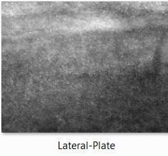
Lateral plate: name of a tissue thickening along the sides of the body, which will, in particular, become the limbs.
|
|
Series of recent HD movies of chicken embryogenesis, in developmental order.
Click on the image to upload the complete film in gif format
Click on the link underneath the image to upload a gif animation of the reduced film scaled by (1/3)x(1/3) in X-Y
Click underneath to download the complete video in .avi
The best movies are indicated by the "three embryos"  tag tag
|

Le męme réduit (1/3)x(1/3)
Vidéo du film complet (58Mo)
|

Le męme réduit (1/3)x(1/3)
Vidéo du film complet (113Mo)
|
Le męme réduit (1/3)x(1/3)
Vidéo du film complet (113Mo)
|

Le męme réduit (1/3)x(1/3)
Vidéo du film complet (300Mo)
|
Le męme réduit (1/3)x(1/3)
Vidéo du film complet (32Mo)
|
Le męme réduit (1/3)x(1/3)
Vidéo du film complet (53Mo)
|
Le męme réduit (1/3)x(1/3)
Vidéo du film complet (11Mo)
|

Le męme réduit (1/3)x(1/3)
Vidéo du film complet (190Mo)
|
Le męme réduit (1/3)x(1/3)
Vidéo du film complet (299Mo)
|
Le męme réduit (1/3)x(1/3)
Vidéo du film complet (98Mo)
|

Le męme réduit (1/3)x(1/3)
Vidéo du film complet (167Mo)
|

Le męme réduit (1/3)x(1/3)
Vidéo du film complet (39Mo)
|
Le męme réduit (1/3)x(1/3)
Vidéo du film complet (85Mo)
|
Le męme réduit (1/3)x(1/3)
Vidéo du film complet (200Mo)
|
Le męme réduit (1/3)x(1/3)
Vidéo du film complet (100Mo)
|

Le męme réduit (1/3)x(1/3)
Vidéo du film complet (53Mo)
|

Le męme réduit (1/3)x(1/3)
Vidéo du film complet (32Mo)
|

Le męme réduit (1/3)x(1/3)
Vidéo du film complet (117Mo)
|

Le męme réduit (1/3)x(1/3)
Vidéo du film complet (91Mo)
|

Le męme réduit (1/3)x(1/3)
Vidéo du film complet (125Mo)
|
Le męme réduit (1/3)x(1/3)
Vidéo du film complet (117Mo)
|

Le męme réduit (1/3)x(1/3)
Vidéo du film complet (142Mo)
|

Le męme réduit (1/3)x(1/3)
Vidéo du film complet (99Mo)
|

Le męme réduit (1/3)x(1/3)
Vidéo du film complet (377Mo)
|
|
Series of older movies of chicken embryogenesis, less resolved, roughly in developmental order
|


|
|
|

|
|
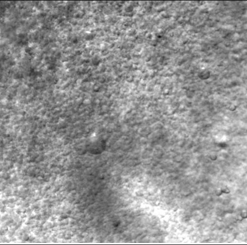
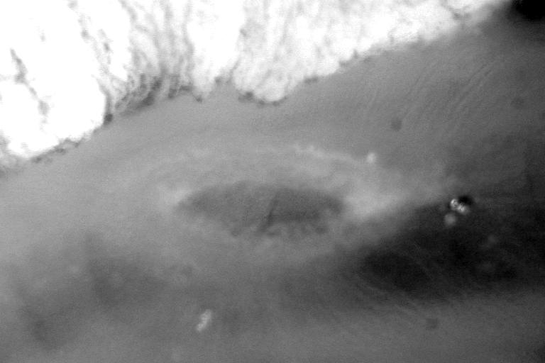
Stage : Initial rotatory movements of the blastula.
Magnification : X5.
Camera: HD 1064x768.
Remarks :The top left film movie shows a global view of the blastula with cellular resolution.
The cells at the periphery are larger than the cells located in the center of the blastula.
A movie was generated for both sides. THis one shows the "underneath view" of the blastula.
One may remark the convection of the larger cells towards the inside of the blastula.
The other movies show another embryo, and above all, a magnified view
of the area of the bigger cells, and the area of the smaller cells.
Personal remark : Emb 506.
|
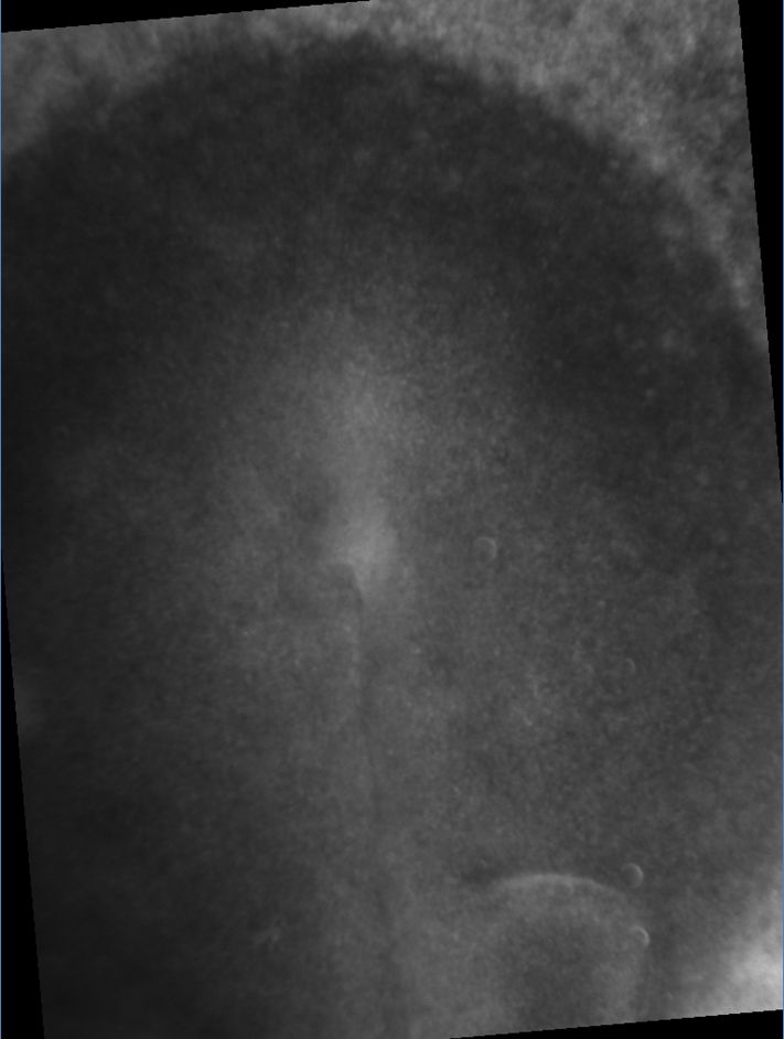
Stage : Initial rotatory movements of the blastula.
Magnification : X4.
Camera: Watec B&W, 768x512 pixels.
Remarks of general interest : This movie
shows a wide field view of the exact moment when the primitive streak opens. It opens in both anterior and posterior directions.
On the edge, the traction forces unstick the blastula from the vitelline membrane, but this seems to have little effect.
Personal remarks : Emb 196.
|
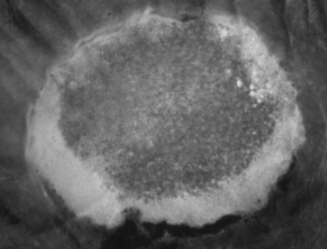
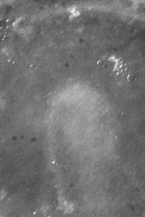
Stage : Initial rotatory movements of the blastula.
Magnification : X4.
Camera: Watec B&W, 768x512 pixels.
General remarks : This movie
shows a wide field view of the blastula.
One may observe here the beginning of the caudal elongation, which is quadrupolar, under the advection of the tissues in the flow.
Personal remarks : Emb 135.
|
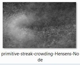
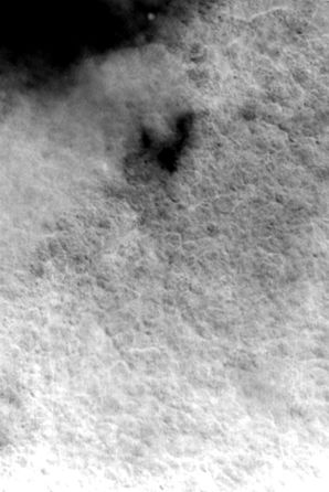
Stage :End of opening of the primitive streak, and engulfment of the top layer.
Magnification : X4.
Camera: Watec B&W, HD1064x768 pixels.
Remarks of general interest : This movie
shows a wide field view of the formation of the primitive streak (as seen from underneath), and the involution of the ectoderm in the streak.
Personal remarks : Emb 505.
|

|
|
|
|
|
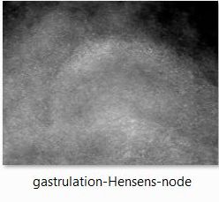
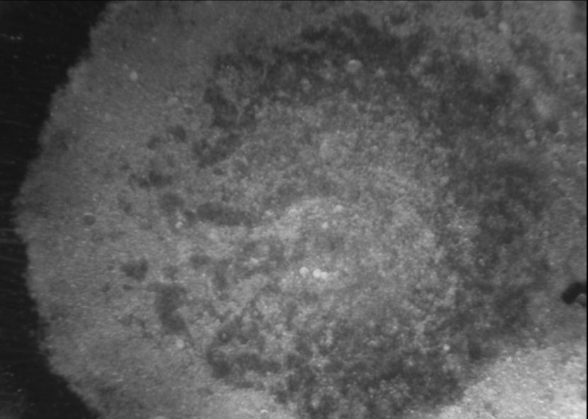
Stage : Gastrulation.
Magnification : X3.
Camera: Watec B&W, 768x512 pixels.
Remarks of general interest : This movie shows that the front of mesodermal cells is pushing away
the gel present inside the embryonic cavity
(in between ectoderm and endoderm).
Personal remarks : Emb 564-1.Almost complete film of mesodermal migration
|
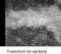
Stage : Gastrulation.
Magnification : X3.
Camera: Watec B&W, 768x512 pixels.
Remarks of general interest : This movie
shows the beginning of mesoderm invagination,
as seen from underneath. One distinguishes clearly the progression of the front of mesoderm.
Personal remarks : Emb 156.
|
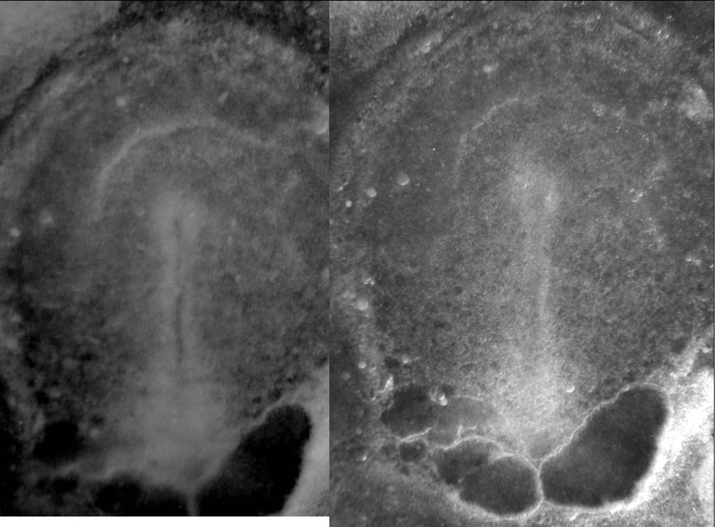
Stage : Gastrulation : End of the opening of the primitive streak and injection of the mesoderm.
Magnification : X3.
Cameras: Watec B&W, 768x512 pixels + Marlin 1064x768.
Remarks of general interest : This movie shows
a wide field view of the embryo during mesoderm injection.
This shows actually a dual dorso-ventral view of the same embryo put on a glass slide. One images very clearly the front of mesoderm.
Personal remarks : Emb TM4, this embryo was generated with pupils from the collčge Thomas Mann.
|
Stage : Gastrulation: End of mesoderm injection.
Magnification : X3.
Camera: Watec B&W, 768x512 pixels.
Remarks of general interest : This movie
shows a wide field view of the injection of the mesoderm, but as seen across the ectoderm.
One distinguishes the frontier
of the mesoderm which is moving forward progressively.
As the mesoderm pulls itself forward on the ectoderm, a curvature (bending) of the ectoderm-mesoderm sandwich is evidenced.
Personal remarks : Emb 71.
|
|
|
|
|
|
Stage : Gastrulation.
Magnification : X1.
Camera: Watec B&W, 768x512 pixels.
Wide field view of a gastrulating blastula.
Personal remarks : Emb 567-1, prepared with Ryan.
|
Stage : Gastrulation.
Magnification : X2.
Camera: Watec B&W, 768x512 pixels.
Same embryo as previous one. A closer look shows a front of cells moving away
a central elongated hole (the primitive streak).
Personal remarks : Emb 567 (avec Ryan).
|
Stage : Gastrulation : End of opening of the primitive streak and injection of the mesoderm.
Magnification : X6,3.
Camera: Watec B&W, 768x512 pixels.
Same embryo as previous one. A close up view of the front, as observed from atop. The film
shows the movement of cells underneath, as seen across the ectodem.
Personal remarks : Emb 567 (with Ryan).
|
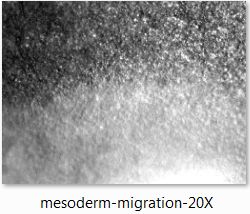
Stage : Gastrulation.
Magnification : X20.
Camera: Watec B&W, 768x512 pixels.
Remark: Very magnified time-lapse film x20 showing the front of cells (mesenchymal cells, fibroblast like)
moving forward (more and better movies to come on this website soon).
Personal remarks : Emb 567 (with Ryan).
|
|
|

|

|
|
|
Stage : Gastrulation.
Magnification : X00.
Camera: Watec B&W, 768x512 pixels.
|
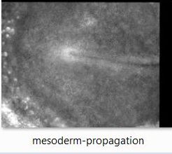
Stage : Gastrulation.
Magnification : X3.
Camera: Watec B&W, 768x512 pixels.
This movie
shows a wide field movie in grazing illumination of the front of mesoderm
springing out of the primitive streak: Emb 572-ZE (with Ryan).
|
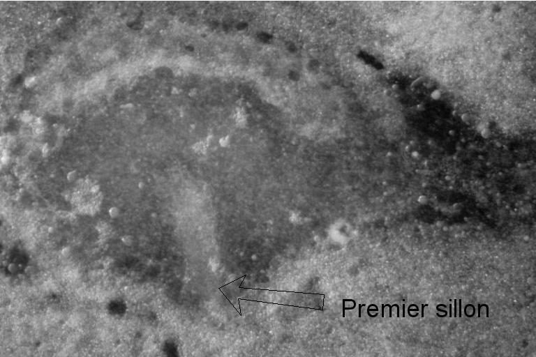
Stage : Gastrulation : Fin d'ouverture du premier sillon et injection du mésoderme.
Magnification : X8.
Camera: Watec B&W, 768x512 pixels.
Same embryo as previous one : the front is seen in close up view, from underneath.
The movie shows the movement of the cells underneath
which are springing inside the primitive streak.
Personal remarks : Emb 567 (with Ryan).
|
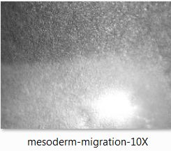
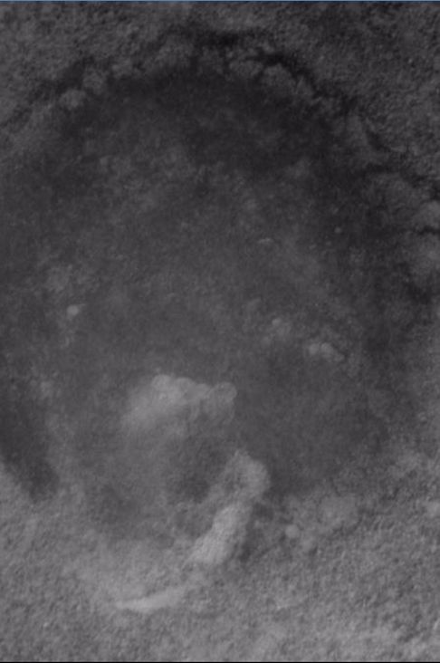
Stage : Gastrulation.
Magnification : X10.
Camera: Watec B&W, 768x512 pixels.
Remark: Close up view (X10) showing the flux of cells springing inside the primitive streak.
(see film).
Personal remarks : Emb 564 (with Ryan).
|
|

|

|
|
|
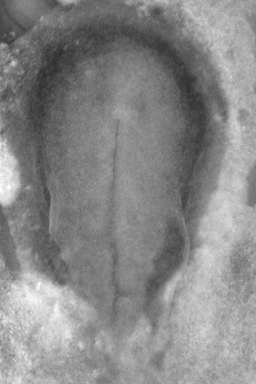
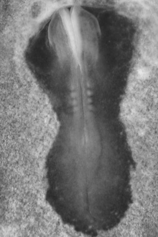
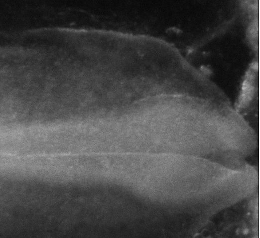
Stage : End of gastrulation X4, beginning of chord formation.
Camera: Watec B&W, 768x512 pixels.
Remarks of general interest :
Traction movement of the mesoderm upon the ectoderm, which provokes a stretch and an antero-posterior fold
(beginning of neurulation).
(The film ).
Personal remarks : Emb 557. By the end the embryo is a bit crippled
Extended remarks
|
Stage :End of the extension of the primitive streak, and beginning of the recession.
Magnification : X2,5.
Camera: Watec B&W, 768x512 pixels.
Remarks of general interest : Here a visible film of the embryo to the left, at magnification X4
film also visible of the embryo to the right at magnificationt X3
(quite spectacular; the entire movie=54Mb)
Personal remarks : Emb 64, used; Emb201 (shown in course with Jean-Pierre).
|
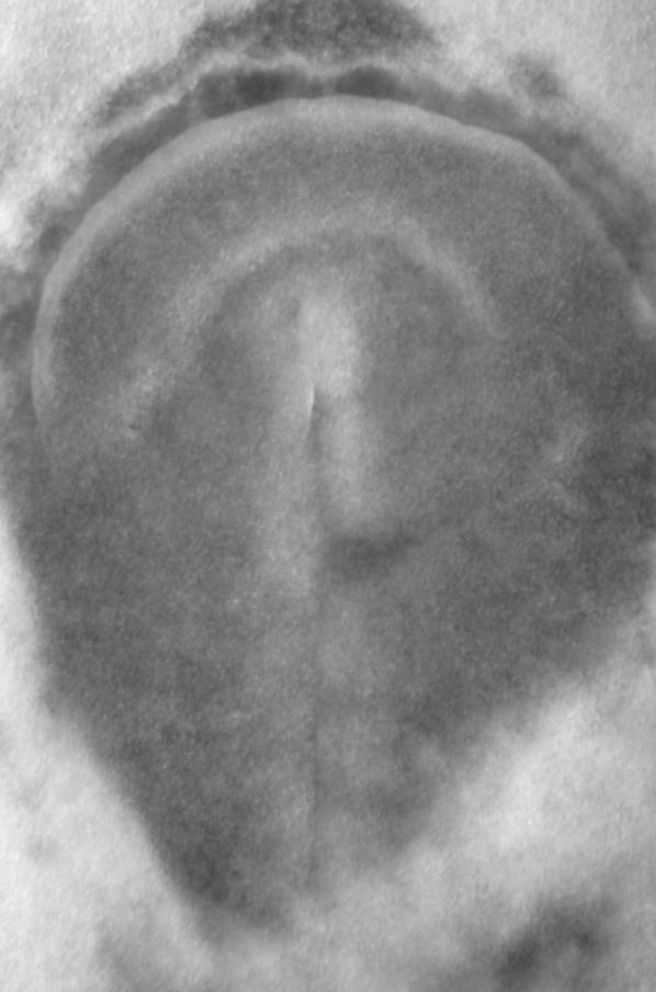
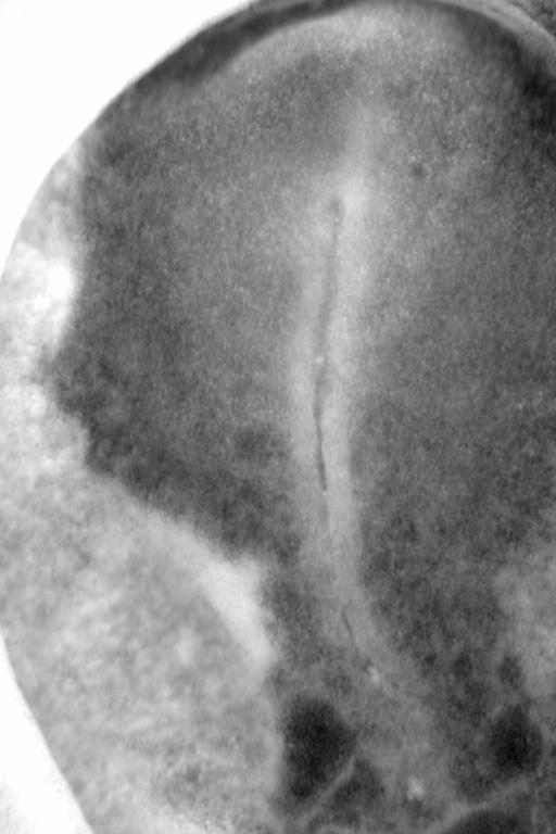
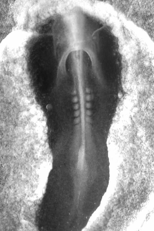
Stage : Early appearance and extension of the primitive streak.
Magnification : X3.
Camera: Watec B&W, 768x512 pixels.
Remarks of general interest :
A film in close up view from atop (ectodermal side= showing how
the primitive streak closes, and the beginning of the extension of the blastula.
Personal remarks : Emb 157. Used for the article in biosystems
Extended remarks
|
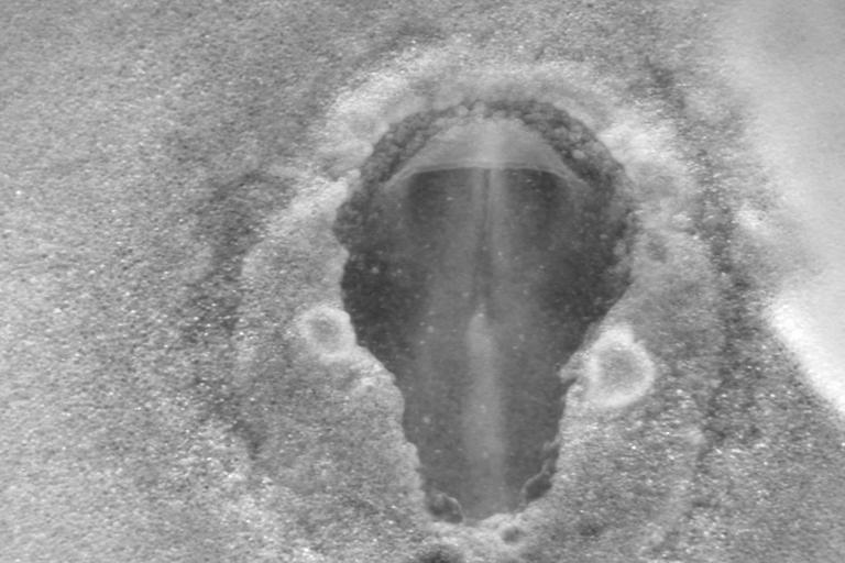
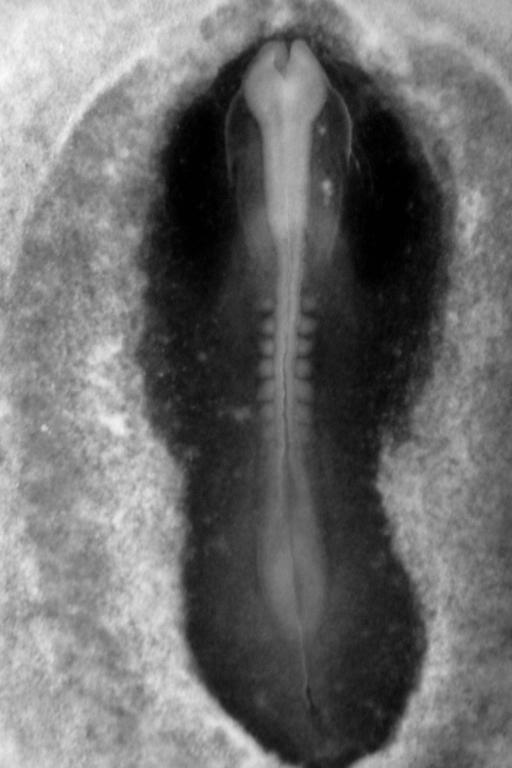
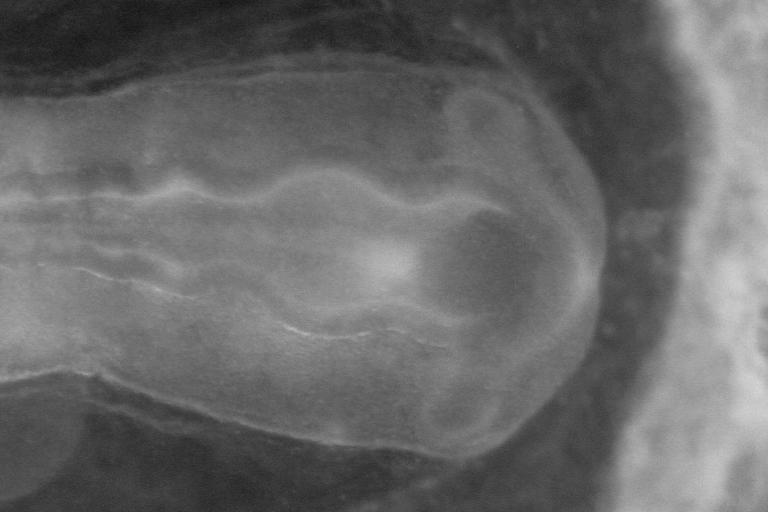
Stage : Recession of the primitive streak. Formation of the cardiac fold. Early
signs of the chorioninc fold, early signs of neural folds.
Magnification : X3,2;.
Camera: Watec B&W, 768x512 pixels.
Remarks of general interest : Formidable film of the extension of the doral folds
and of formation of vortex-like windings (quadrupolar stretch)
Remark personnelle : "perfect" embryon.
|
|
|

|
|
|
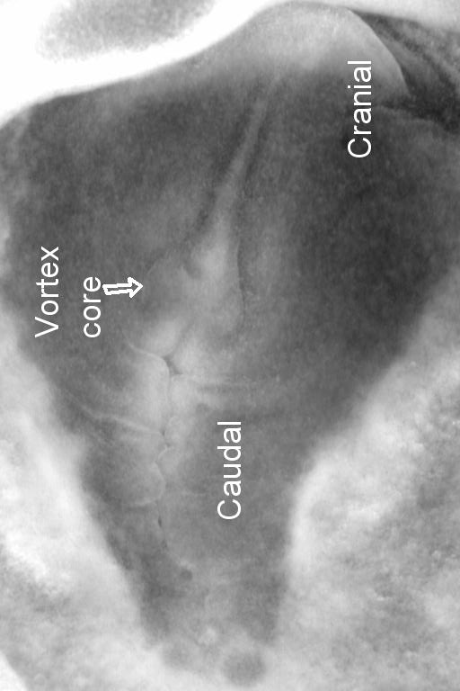
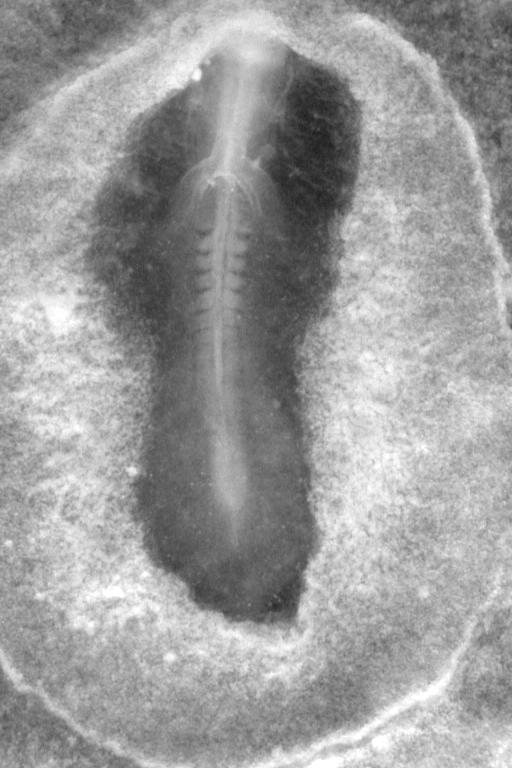
Stage : Moment of shear at the ectoderm surface, by the end of gastrulation, as seen from underneath.
The shear generates the gastro-cardiac fold.
Magnification : X3,2.
Camera: Watec B&W, 768x512 pixels.
Remarks of general interest :
This Film shows the extension of the (futur) dorsal area, as seen from the ventral side. One sees
the formation of vortices, and the early folding of the gastro-cardiac pocket.
Personal remarks : Emb 40 (x3,2), utilisé (x2,25); Emb 129; Emb 196.
Extended remarks
|
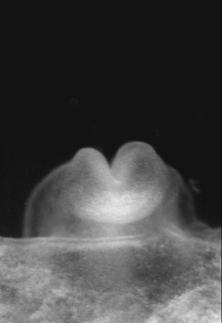
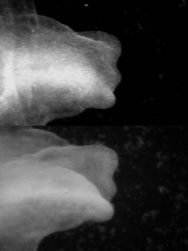
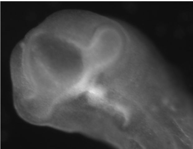
Stage : Neurulation.
Magnification : X4.
Camera: Watec B&W, 768x512 pixels. This
film shows the formation of the folds in the head, in frontal view.
Remarks of general interest : Embryon ML12 generated with Manon.
|
Stage : Neurulation (formation of the brain and of the optic cupulas).
Magnification : X5.
Camera: Watec B&W, 768x512 pixels.
Remarks of general interest :
A film in close up view and in 3/4 orientation showing the entire neurulation
of the anterior part of the embryo.
Personal remarks : Emb ML10, generated with Manon.
Extended remarks
|
Stage : Neurulation (formation of the brain and of the optic cupulas).
Magnification : X4.
Camera: Watec B&W, 768x512 pixels.
Remarks of general interest : exceptional polar view of the head during neurulation.
The film in close up view shows a polar-frontal view of the entire neurulation of the anterior part of the embryo.
Personal remarks : Emb ML4 et ML5, generated with Manon.
Extended remarks
|
 |


|

|

|
|
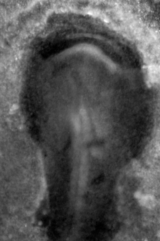
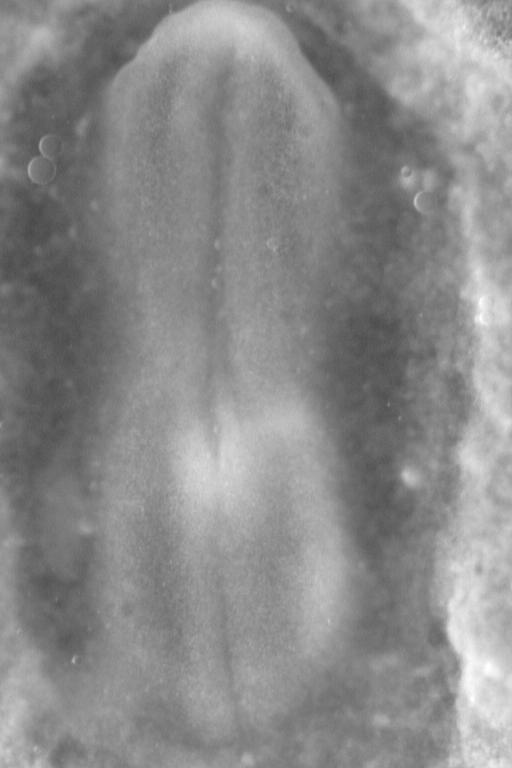
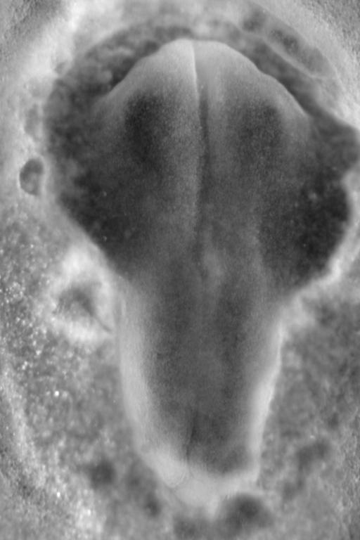
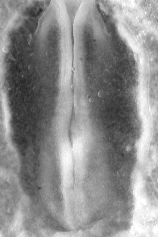
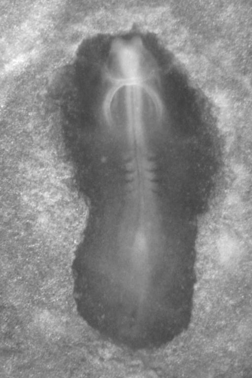
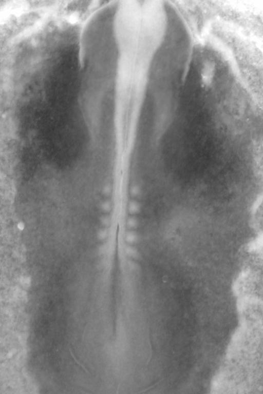
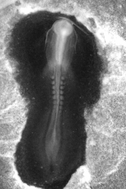
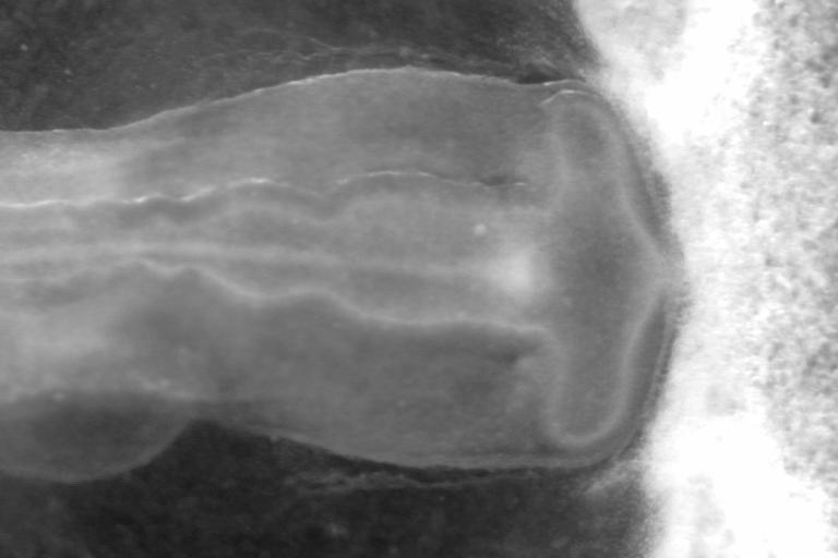
Stage : End of primitive streak.
Magnification : X3.8.
Camera: Watec B&W, 768x512 pixels.
General remarks : Watch the movie.
You can guess the cardiac fold at the top, which forms a crescent. You see the formation of the chord
(primitive dorsal axis of all vertebrates and also of cephalochordates).
Personal tags: Emb 39, utilisé.
Extended remarks
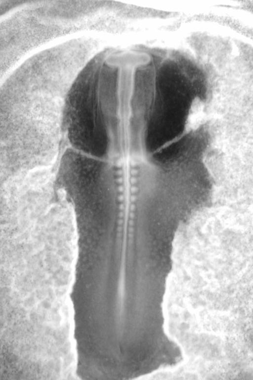
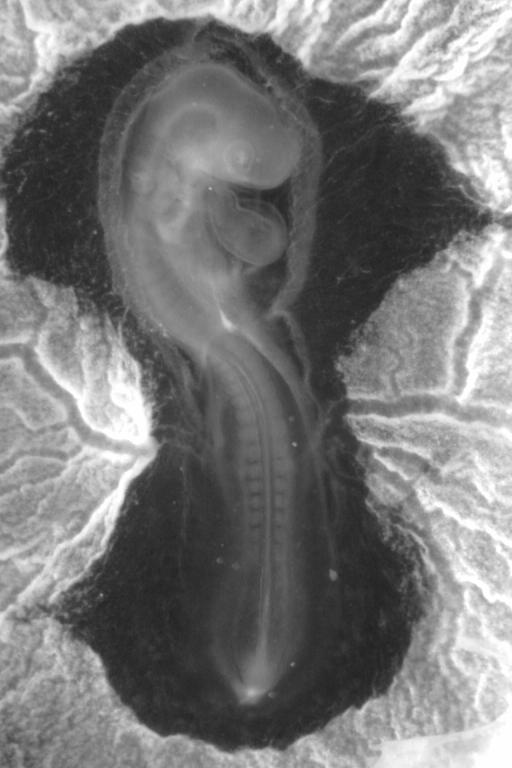
Magnification : X3.8.
Camera: Watec B&W, 768x512 pixels.
General remarks :Watch the movie.
You see the extension of the posterior area, and formation of the pelvic winding.
It is the same embryo as the one before.
Personal tags: Emb 39 used.
Extended remarks
|
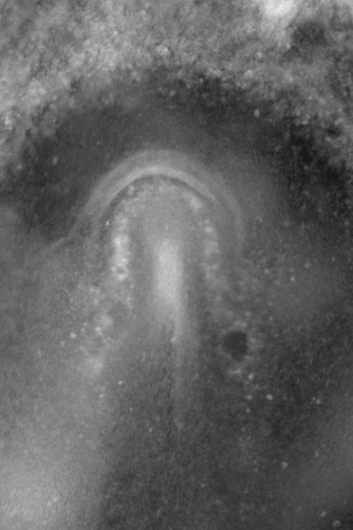
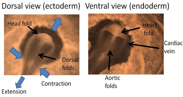
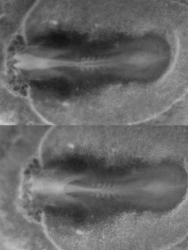
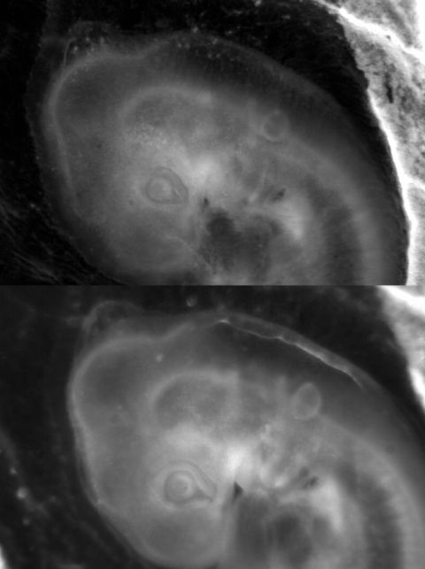
Stage : Roll up of the neural crest and formation of the gastro-cardiac pocket (embryo #145 X3, embryo#165X2).
Cameras: Watec B&W, 768x512 pixels.
Remark : Formidable double movie
of the formation of the embryo, seen simultaneously, on the very same embryo, in dorsal and ventral view.
There is some noise due to a Sync. problem between the two cameras.
Please note the flatenning along the median axis, the recirculation around the pelvic and scapular area.
The physical nature of the process is very obvious.
Second double movie
of embryo formation showing a double view, from atop, and underneath, on the very same embryo.
|
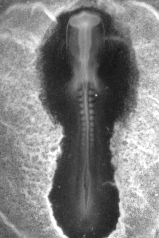
Left; Stage : beginning of neural crest roll-up.
Magnification : X5.
Camera: Watec B&W, 768x512 pixels.
General remarks :Watch the movie.
You see the beginning of the roll up of the dorsal area.
Personal tags: Emb 110.
Extended remarks
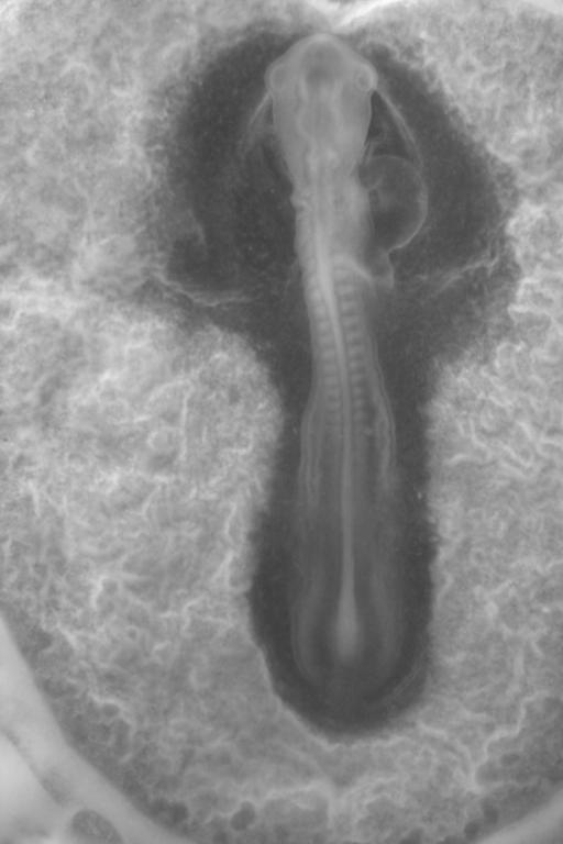
Right; Stage : beginning of neural crest roll-up.
Magnification : X3.5.
Camera: Watec B&W, 768x512 pixels.
General remarks :Watch the movie.
Wonderful movie showing the entire roll-up of the dorsal area.
The formation of the brain folds is very well seen.
Personal tags: Emb 36. Used.
|
Film of the appearance of the heart-gut fold. At the beginning of the movie, the blastula is flat. The extension shears the surfae and the underneath layers.
The buckling creates a fold which progresively constricts. Unfortunately some yolk was left on the ventral side. The movie is quite long, therefore it was reduced to 0.33X, and only even slices are shown.
The full movie is 768x1024pxls, and contains about 600 plates.
|
|
 |
 |
|
|
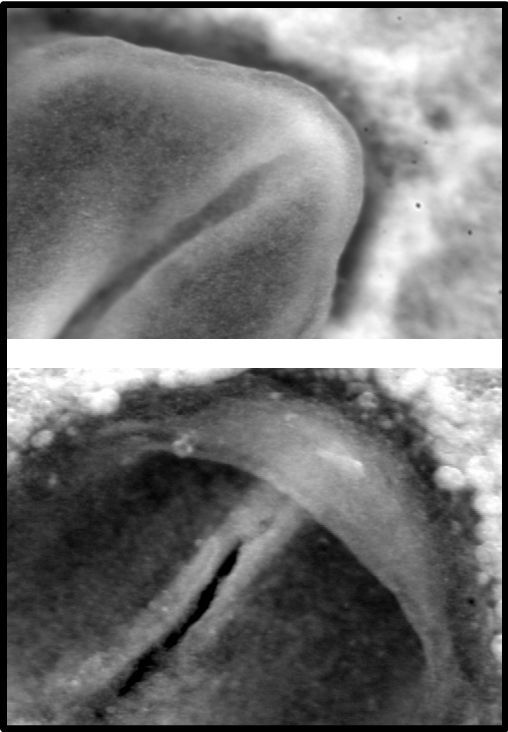
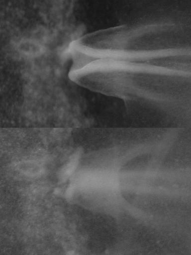
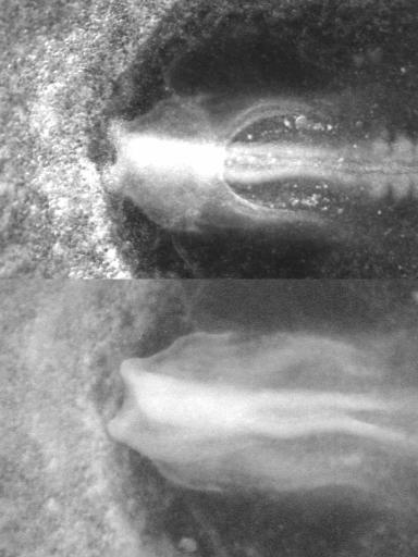
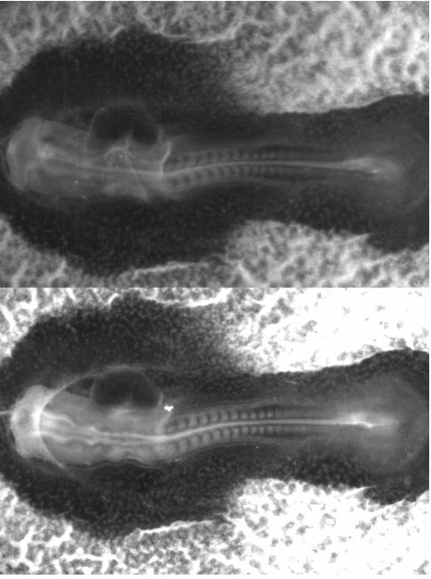
Stage : Left: the gut-heart fold, as seen from underneath,
Right : formation of the cephalic fold on the same embryo.
Magnification : X3.5 for the bottom view X4 for the top view
Camera: Watec B&W, 768x512 pixels.
General remarks : the underneath view is very clear, and enlightning.
Personal tags: Emb XX.
Extended remarks |
Stage : 0 somites
Magnification : X4.
Camera: Watec B&W, 768x512 pixels.
General remarks : Double simultaneous view at the moment of formation of the
cardiac pocket.
Personal tags: Emb 10bis.
Extended remarks
|
Stage : 0 somites
Magnification : X4.
Camera: Watec B&W, 768x512 pixels.
General remarks : Other embryo, double simultaneous view just before the moment of formation of the
cardiac pocket.
Personal tags: Emb XX.
Extended remarks
|
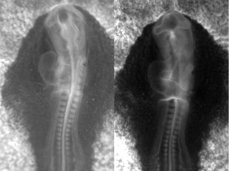
Stage : To the left, exact moment when the neural crests from left and right contact and merge. To the right, magnification of the head area.
Magnification : X4. To the right
Camera: Watec B&W, 768x512 pixels.
General remarks : Watch the movie of the left image.
We witness the collision of the two halves of the embryo (left and right) along the median axis.
The embryo stretches bi-directionnaly.
The picture to the right corresponds to : a movie.
showing the fate of the cephalic area, as it is pulled down by the cardio-gut pocket.
Personal tags: Emb 36 and Emb 168.
Extended remarks
|
 |
|
|
 |
|
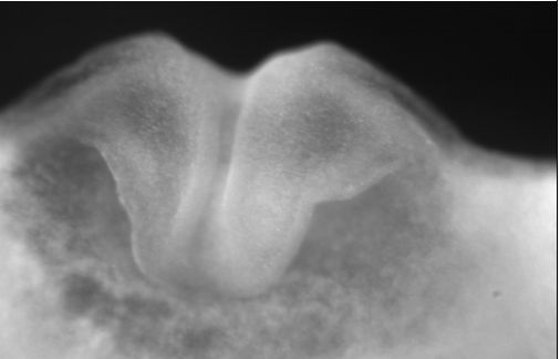
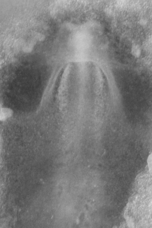
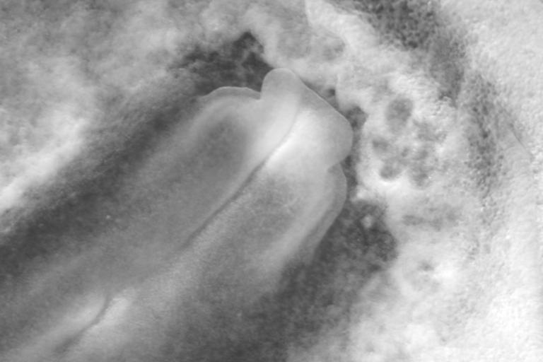
Stage : 3 somites.
Magnification : X3.2.
Camera: Watec B&W, 768x512 pixels.
General remarks :Watch the movie. Beginning of heart formation :
a very simple fold in the shape of a hood.
Personal tags: Emb 102.
Extended remarks
|
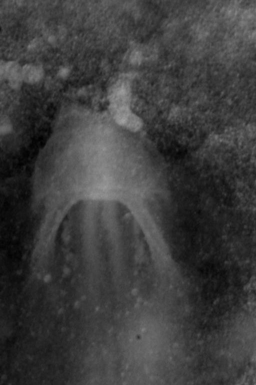
Stage : 3 somite pairs.
Magnification : X3.2.
Camera: Watec B&W, 768x512 pixels.
General remarks : Watch the movie. Continuation of cardiac constriction
See also Almost complete movie of the formation of the heart by contraction of the cardiac fold.
Personal tags: Emb 101.Emb 178
Extended remarks
|
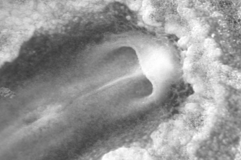
Stage : 4 somite pairs.
Magnification : X1.6.
Camera: Watec B&W, 768x512 pixels.
General remarks : Watch the movie.
Formation of the larger vessels of the heart (edges of the hood-fold).
In the second movie one sees the beginning of the looping of the cardiac tubes.Second film
Personal tags: Emb 103 (it is the same embryo).
Extended remarks
|
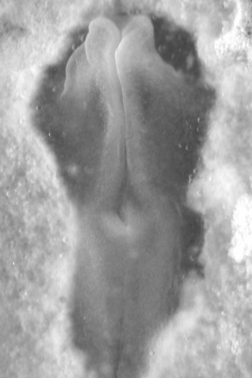
Stage : Beginning of head evagination and swelling.
Magnification : X4.
Camera: Watec B&W, 768x512 pixels.
General remarks :The frontal folds of the head spread laterally (some sort of a trail), as the median axis pushes forward.
Personal tags: Emb XX.
Extended remarks
|
 |
 |
|
|
|
Stage : Top : The head passes over the cardiac fold, dorsal view.
Magnification : X4.
Camera: Watec B&W, 768x512 pixels.
General remarks :
Personal tags: Emb 23.
Extended remarks
Stage : Botom : The head passes over the cardiac , ventral view
Magnification : X4.
Camera: Watec B&W, 768x512 pixels.
General remarks :It is the same embryo. The fold located anteriorily will be the heart
Personal tags: Emb 23.
Extended remarks
|
Magnification : X2.2.
Camera: Watec B&W, 768x512 pixels.
General remarks : One sees very well that the head has passed over the blastula plane like a finger of a glove.
Personal tags: Emb 26.
Extended remarks
|
Stage : 3-5 somites.
Magnification : X3 and X2.5
Camera: Watec B&W, 768x512 pixels.
General remarks : visible movies dorsal view, and
ventral view
One sees the swelling of the neural folds, the body extension, and the pelvic winding.
On the ventral side, a large part of heart formation is seen (collapse of folds forming the heart structure).
Personal tags: Emb 28.
Extended remarks
|
Stage : 5 somites.
Magnification : X1.9; X4; X6.3
Camera: Watec B&W, 768x512 pixels.
General remarks : Left : double movie of the same embryo seen simultaneously in dorsal and ventral view.
The movie.
Middle : double movie of the same embryo seen simultaneously in dorsal and ventral view (X4).
The movie.
Right : higher resolution double movie of the same embryo seen simultaneously in dorsal and ventral view.
The movie.
One sees the collapse of the anterior part of the brain, likely under the stretch
of the cardiac pocket.The cardiac fold is quite visible, as it constricts and descends
Extended remarks
|
|

|

|
 |
|
Stage : 6 and 7 somites pairs.
Magnification : X2.4.
Camera: Watec B&W, 768x512 pixels.
General remarks : Watch the movie
Swelling of the head, body extension, winding of the pelvic area.
Next a movie showing the closure of the nasal pit, and the
start of the evagination of the eye primordium.
Personal tags: Emb 70, Emb M0.
Extended remarks
|
Stage : 7 somites pairs.
Magnification : X1.6.
Camera: Watec B&W, 768x512 pixels.
General remarks :
Watch the movie. Ventral view of the embryo, the cardiac fold is very well seen.
Next, a film.
of the lateral evagination of the eye primordium, under the forward push of the median axis.
Along the neural crest one sees the migration of neural cells which move away from the midline
Personal tags: Emb 20. Emb 55
Extended remarks
|
Stage : 8 somites pairs.
Magnification : X2.
Camera: Watec B&W, 768x512 pixels.
General remarks : Left static image X1.6 : You see how the head pushes over the blastula and creates a fold "ahead of the head".
The vitelline veins are hardly visible, on the sides of the cardiac arches.
Right : a double dorso/ventral view of development during 4 hours showing the evagination of the eye cups, the looping of the heart etc.
Personal tags: Emb 56. Emb175.
Extended remarks
|
Stage : 9 and 10 somites pairs.
Magnification : X1.6.
Camera: Watec B&W, 768x512 pixels.
General remarks : Beginning of the evagination of the eyes cupula.
The head extension contributes to pushing the eye field sideways.
Personal tags: Emb 21 et 53.
Extended remarks
|


|
 |
|
 |
|
Stage : 12 somites.
Magnification : X1.6.
Camera: Watec B&W, 768x512 pixels.
General remarks : Left Ventral view, the heart is well visible.
Right double ventral and dorsal view of the same embryo.
The eye cupulas start to bend posteriorily on the head of the sides (sort of trail).
The chorio-amniotic fold starts to pass over the head.
The movie shows 4 hours of development at this stage.
Watch the inflation of the brain vesicles, and the growth of the heart. The somites drif anteriorily.
Personal tags: Emb 67. Emb 176
Extended remarks
|
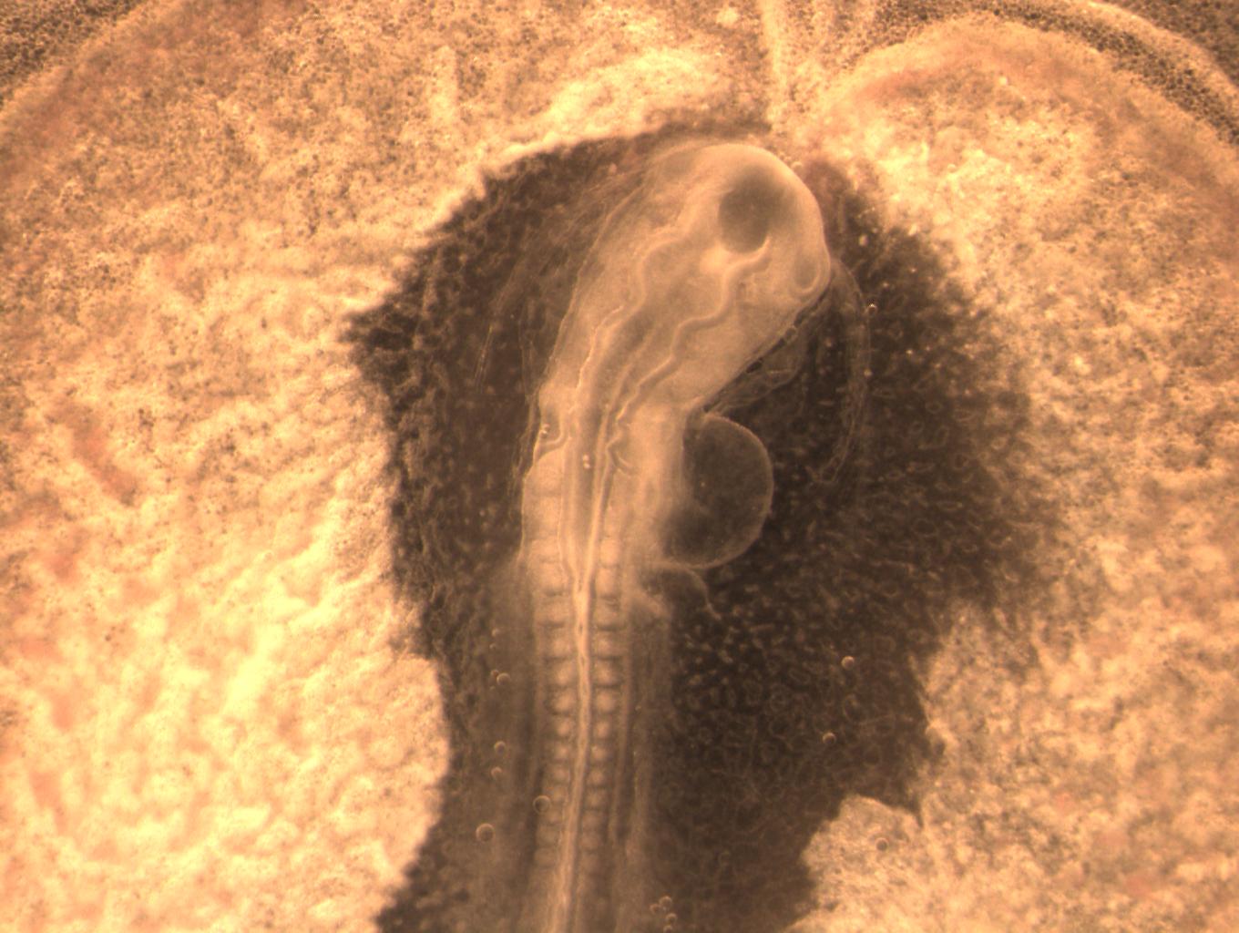
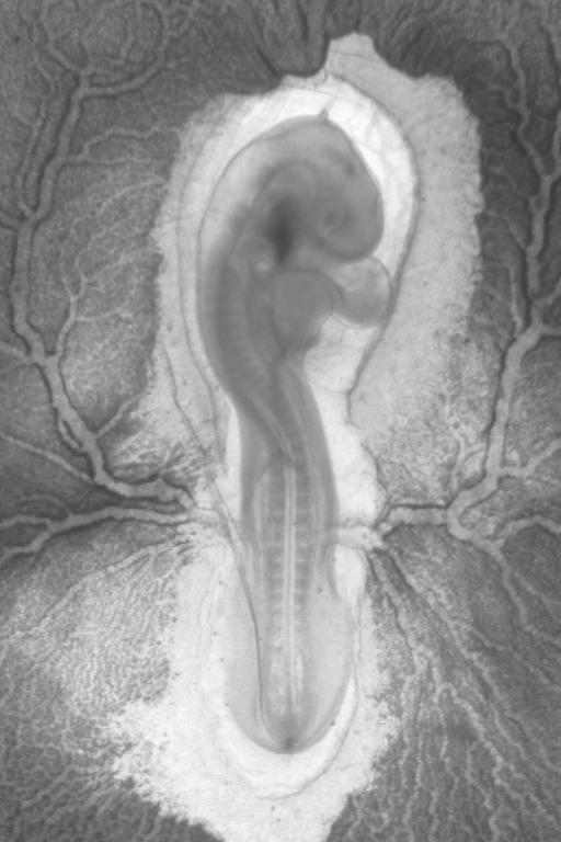
Stage : 14 somites, formation of the chorion-amniotic fold.
Magnification : X1.25.
Camera: Watec B&W, 768x512 pixels.
General remarks : the head movie.
One may see the beginning of the evaginations of the eyes in the trail of the median axis.
In the Movie you also see the formation of a bumpy fold "ahead of the head", which is eventually the chorion.
The brain vesicles are starting to dilate. The domains of the limbs are well visible.
Personal tags: film=Emb 110.
Extended remarks
|
Stage : 16 somites. The chorionic fold is passing over the head, the eyes start to form.
Magnification : X2.2.
Camera: Scion Colour, high definition.
General remarks : another film of the head.
One clearly sees how the eyes wind closer to the sides of the future brain (trail effect).
The fold of the chorion is quite visible as it forms a hood over the head.
The heart is visible.
Personal tags: Dossier Embryons complets.
Extended remarks
|
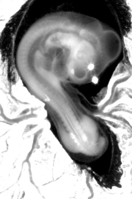
Stage : 22 somites.
Magnification : X0.71.
Camera: Watec B&W, 768x512 pixels.
General remarks : A well recognizable head. The closure of the chorio-amniotic bag is in progress.
Personal tags: Emb 52.
Extended remarks
|
 |

|
 |
|
|
Magnification : X1,6.
Camera: Watec B&W, 768x512 pixels on both sides. A Leica microscope and an optem zoom (inverted).
Remarks of general interest : Simultaneous dula dorso-ventral image
of an embryo during evagination of optic cups.
One sees the chorionic fold passing over the head. One sees the morphogenesis of the heart.
Film visible ,
Personal remarks : Emb 176.
Extended remarks
|
Stage : Eye formation.
Magnification : X6.
Camera: Watec B&W, 768x512 pixels.
Remarks of general interest : Film of the formation of the eyes as seen in frontal view.
One distinguishes the flatening of the eyes on either sides, andthe triggering of the
lens and of the optic chamber (see eye on the right in the field).
Personal remarks : EmbML18.
Similar kind of film of the formation of the eyes as seen in frontal view.
One distinguishes the flatenning on the sides, and the triggering of the lens and of the optic chamber. (see eye at top left in the image)
Personal remarks : EmbML21.
Extended remarks
|
Stage : neural flexure
Magnification : X4.
Camera: Watec B&W, 768x512 pixels.
Remarks of general interest : Double 3/4 imaging in both dorsal and ventral view of the same embryo.
film visible.
One distinguishes the evagination of the eyes and the formation of the mouth fold.
The optic cupula flattens as it moves up against the ectoderm.
The brain starts to dilate.
Personal remarks : film=Emb 428.
Extended remarks
|
Magnification : X1,25.
Camera: Watec B&W, 768x512 pixels on both sides. An upright Leica microscope and an iverted Optem zoom.
Remarks of general interest : Dual simultaneous dorso-ventral image of an embryo during evagination of the optic cupulas.
One sees the progression of the fold of the chorion over the head.
Ine sees the formation of the heart and of the omphalomesenteric veins (the navel), which move "down" towards the hyperbolic point area.
Film visible ,
Personal remarks : Emb 202.
Extended remarks
|
 |
 |
 |
 |
|
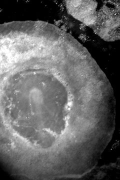
Stage : Formation of the optic cupulas.
Magnification : X6.
Camera: Watec NetB, 768x512 pixels.
remarks of general interestThis film shows the early stage of cupulas evaginations
Remarque personnelle: Emb510
Extended remarks
|
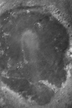
Stage : Same as previous one
Magnification : X6.
Camera: Watec NetB 768x512.
remarks of general interestThis film shows the formation of the optic cupulas.
Personal remarks : Emb517.
Extended remarks
|
Stage : extension of evaginations
Magnification : X8.
Camera: Marlin HD 1024x768 pixels.
remarks of general interestOne sees the flatenning of the optic cupulas against the ectoderm in a "facing the eye" view.
One remarks that the mouth area is pulling on the head, which winds rostrally, while the brain area (top left fom the eye) is pushing forward.
(The rostral part of the head is hence in a state of shear).
Film visible.
Personal remarks : Emb 555.
Extended remarks
|
Magnification : X2,25.
Camera: Watec NetB, 768x512 pixels.
remarks of general interestDual left and right view of the same embryo with two B&W analog cameras.
The film is visible.
The eyes are formed. One can feel the forward rotation of the encephalon, the lenghtening of the lips, and the formation of the nostrils.
Personal remarks : Emb 430
Extended remarks
|
 |
 |
 |
 |
|
Magnification : X6.
Camera: Marlin HD 1024x768 pixels.
remarks of general interest Lateral evagination of optic cups, beginning of head flexure. 3/4 back view.
Approx. 6hrs of movie.
The Film.
Personal remarks : Emb ML27.
Extended remarks
|
Magnification : X6.
Camera: Marlin NetB, 1024x768 pixels.
remarks of general interest
Beginning of lateral evagination of the optic cups, as seen laterally (profile view). Approx. four hrs of movie.
The movie.
Personal remarks : Emb T4
Extended remarks
|
Stage : 14 somites, formation of the chorionic fold.
Magnification : X5.
Camera: Watec NetB, 768x512 pixels.
Remarks of general interest film of head morphogenesis, prior to diving below the amniotic sac.
Close up view of the evagination of the optic cupulas. One witnesses a discontinuity. The brain starts to dilate distinctly.
Remarque personnelle: EmbTM8
Extended remarks
|
Stage :
Magnification : x5.
Camera: Watec NetB 768x512.
remarks of general interest film of had development seen from a 3/4 frontal view during eye formation.
One sees clearly how the eyes move along the sides of the future brain (sort of wake effect).
Personal remarks : EmbML14.
Extended remarks
|
 |
 |
 |
 |
|
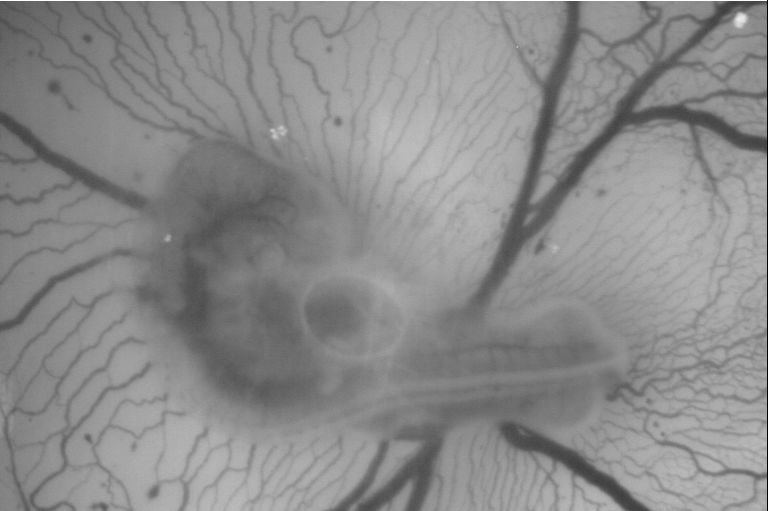
Stage : 28 somites.
Magnification : X0.71.
Camera: Watec B&W, 768x512 pixels.
General remarks : Transmission view, the vessels of the yolk sac are visible
Personal tags: Emb 81.
Extended remarks
|
Stage : approx. 32 somites.
Magnification : X0.71.
Camera: Watec B&W, 768x512 pixels.
General remarks : Watch the movie.Film of the closure of the chorio-amniotic bag.
It closes around a singularity (a hole) which remains as a scar on the bag. It is some sort of a navel, with no umbilical chord accross it.
Also, a close up view showing the closure, and the ripples created by the constriction of the hole
(much similar to wound healing).
Watch the movie.
Personal tags: Utilisé pour article CRAS.
Extended remarks
|
Stage : approx. 32 somites.
Magnification : X0.71.
Camera: Watec B&W, 768x512 pixels.
General remarks : The limb buds are visible.
Personal tags: Utilisé pour comparaison avec stégosaures.
Extended remarks
|
Stages : diverse stages.
Magnifications : diverse.
Camera: Watec B&W, 768x512 pixels.
General remarks : Snapshots of head formation.
Personal tags:
Extended remarks
|
| Suite des images d'embryons Voir les images
|
Retour à la page de présentation-Back to front page |
|
La citation de la page :
|
"Votre théorie est folle, mais pas assez pour ętre vraie", Niels Bohr.
|


