A few words about capillary regression, a crucial morphogenetic process, and the self-organized formation of veins exactly parallel to arteries.


|
A few words about capillary regression, a crucial morphogenetic process, and the self-organized formation of veins exactly parallel to arteries. |
|||
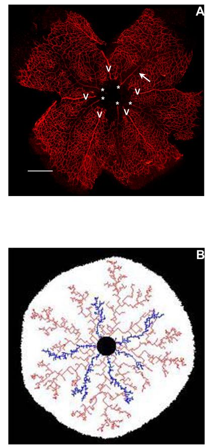
| There exists during vessel morphogenesis a crucial phenomenon, which I call capillary regression. When one watches young arteries, one obsreves that they "detach" spontaneously from the surrounding capillaries. The plate here to the right shows a sambucus nigra staining of endothelial cells in the mouse retina (courtesy Ferdinand Le Noble). One immediately observes that there is some sort of an empty area along the arteries, as if it were devoid of capillaries. (imaging with a Leica fluorescence microscope) | |
||
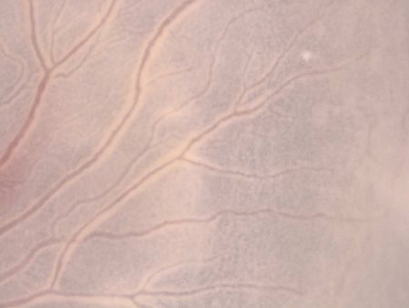
| This peculiar zone, neighbouring the arteries, is actually always visible, in all samples. The plate here to the right shows a direct optical image (Scion corp camera, 12 bits, image taken with an optem Zoomx12). You can guess the presence of some "white" area around the vessels : this whiter area corresponds to a reduced density of capillaries, since red corresponds to presence of blood, hence of vessels. | |
||
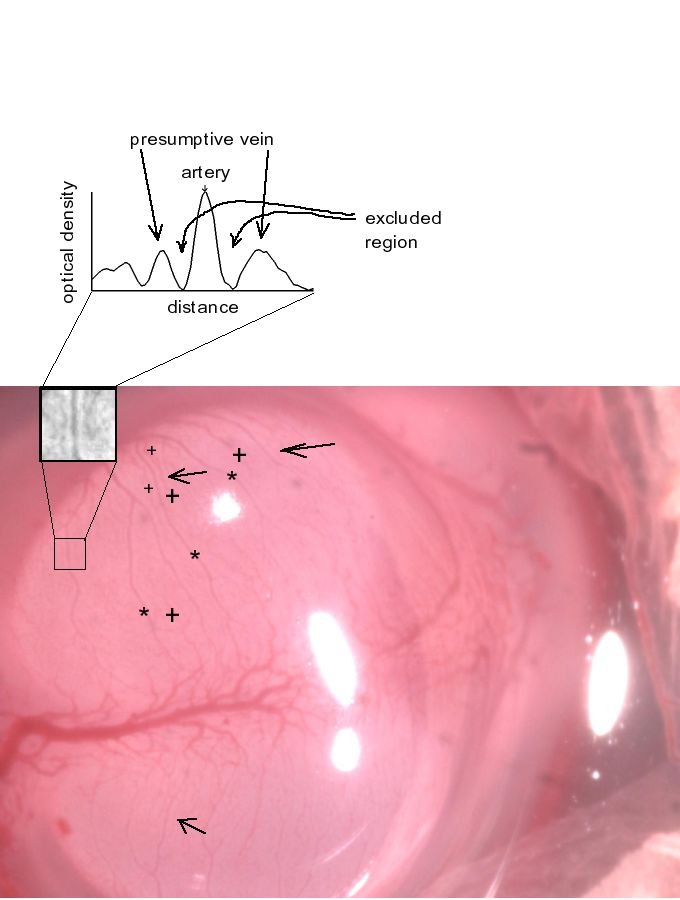
| By integrating the red color, one can show that the presnece of a vessel such as an artery comes together with the absence of capillaries in the neighborhood (at least, a reduction of density). The plate to the right shows a brain vesicle in a chicken embryo, at approx. 6 days of development, in a polar view. When we integrate the red color, we clearly see the depleted zone, and the enhancement somewhat away. This means that formation of an artery comes together with a zone of depletion, close to the artery, and next a zone of enhancement : of what? Of the total volume of cavities through which blood flows. Therefore there is a physical gradient of porosity, which self-organizes the selection or induction of a vein, just nearby an artery (parallel to it, as in the umbilical chord). | |
||
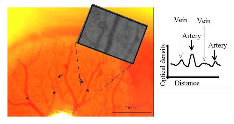
| The same sort of measurement, in a lateral view, on another embryo, a bit younger. (These data belong to Alia Al-Kilani's thesis, and were published in Phys Rev E, see publication list) | |
||
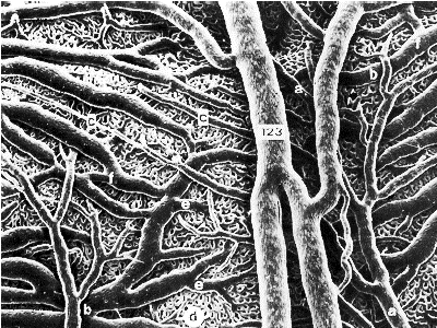
| A few years ago, I put forward the idea that capillary regression was a vital phenomenon, since it makes possible the formation of true vessels, but also because it plays a role in the global pattern formation, by separating arteries and veins, and making possible the direct induction of veins by arteries. Capillary regression makes possible the crossing of arteries and veins, which actually do not "see" each other. To the right, a typical SEM image of a cast of blood vessels (a retina) Photo P. Simoens, Universität Gent, School of veterinary science, with permission. | |
||
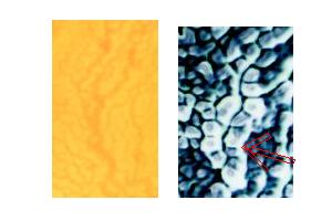
| Together with my students and with Sylvie Lorthois from the Institut de Mécanique des Fluides de Toulouse, we have very finely studied the close neighborhood of arteries. We have shown by shadowgraph, that this area is swollen, around young arteries; and that this swelling participates both in arterial and venous formation. The classical image by binocular microscope does not show any feature, while actually the shadowgraph reveals the detail of stresses and deformation fields (hetre a swelling). | |
||
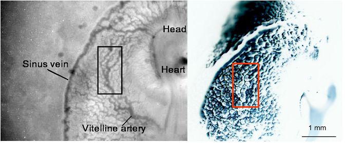
| The very same area as before, seen in a close up view. | |
||
| The swelling induces a detachment of the capillaries, the extra endothelial cells which escaped from that area fuse together further away where they produce a vein parallel to the artery from which they detached. To the left, scheme of the effect of capillary regression. The crushing of capillaries creates a gradient of endthelial cells towards the neighborhood of arteries where a venin forms. | |
||
| We have used Scanning Air Puff Tonometry to measure in vivo the compliance or conversely the stiffness of the areas close to the arteries (the younger ones). Veins appear to form in the softer areas.The flow pattern seems to prune vessels in the more compliant areas. The plate to the right shows measurements of the amplitude of the deformation in response to a compressive air puff. Green curves show the response curve to one shot far away from an artery: the deformation is large. In purple, the response to one shot, just nearby an artery : the response is weak (stiffer material). | |
||
| In vivo image of the tonometer tip, flying over the embryo. This gives a flavour of how the measurements are performed. Quite difficult to be honnest. We get in vivo data about the stiffness of the tissues, with this high resolution tonometer. The embryo proper is oriented vertically in this image. One sees the yolk-sac and the surrounding vessels, wich are just being born. (so to speak). One sees, by the same token, that there exists a lot of roughness locally, which comes togetehr with the valleys and bumps of vessel formation. This is due to the fact that the light is oriented at 45° therefore there exists also a shadowgraphic effect. | |
||
| Images showing the flight over the embryo, when measuring local visco-elastic properties, without contact (although, the air puff is some sort of a contact) The technical details may be found in Physical Review E, Al-Kilani et al. During vertebrate development, arteries exert a morphological control on veins through physical factors 2008); also on this site at publications. | |
||
| Measurement of the stiffness gradient, while scanning perpendicularly to an artery. The very young vein is chosing a path which seems indeed to follow the softest possible path. | |
||
| The quotation of this page : | "Science is built upon facts, just as a house is built with stones, however a heap of stones is not a house nor is heap of facts a science.» Henri Poincaré | ||
| Retour à la page de présentation-Back to front page | |||